医科学専攻 保健学専攻
- Master's Courses
修士課程 - Doctoral Courses
博士課程
Medical Physics医用物理学
STAFF
Professor
-
Gonda, KohsukeProfessor.Ph.D. 権田 幸祐 教授
Other Faculty / Staff
-
Kitamura, Narufumi
Assoc.Prof.Ph.D. 北村 成史 准教授
CONTACT
TEL:+81-22-717-7579
E-MAIL:medphys.tohoku*gmail.com
(「*」を「@」に変換してください)
OUTLINE
疾患の早期診断や治療を効果的に進めるためには、精度の高いイメージング技術が重要です。我々はモデルマウスやヒト組織を対象に研究を行い、血管病変に関与するがん、血栓症、糖尿病、腎症、骨粗鬆症、筋萎縮、認知症など多様な疾患について、その発症や進行に関わる病態機序の解明に取り組んできました。そして得られた知見を、正確で効率的な診断法や治療法の開発につなげることを目標としています。特に、機能性ナノ粒子を利用した高精度かつ高感度なX線CTや蛍光イメージングを開発し、従来技術では困難であった微細な病変の検出を可能にしています。X線吸収は造影剤量に比例し、蛍光強度は励起光に比例するため、両者は定量性の高い可視化手法です。これまでに得られたイメージングデータは病態の理解を深めるだけでなく、新たな診断法や治療法の基盤技術へと発展し、有用な成果を挙げています。さらに近年はナノテラスの放射光(高輝度X線)を活用し、高分解能な病態組織イメージングにも挑戦しています。医用物理学分野では理工医の多様な専門を持つ研究者が集まり、知識や技術を融合させ、協力を大切にしながら真摯に研究を進めています。
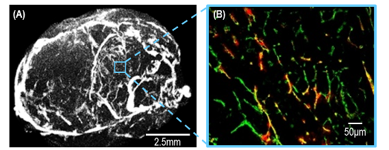
Tumor vessels visualized by X-ray CT (A) and fluorescence (B)
X線CT(A)と蛍光(B)で可視化した腫瘍血管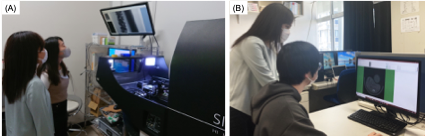
X-ray CT Imaging (A) and analysis (B)
X線CT(A)を用いた画像取得と解析(B)の様子
ARTICLE
Kitamura N et al. Microregional HER2/HER3 density on cancer cells based on multi-point binding detection using nanoparticle-modified cantilever AFM. Colloids Surf. B Biointerfaces 256: 115043, 2025
URL:https://doi.org/10.1016/j.colsurfb.2025.115043
Une N et al. The anti-angiogenic agent lenvatinib induces tumor vessel normalization and enhances radiosensitivity in hepatocellular tumors. Med. Oncol. 38: 60, 2021
URL:https://link.springer.com/article/10.1007/s12032-021-01503-z
Inose T et al. Development of X-ray contrast agents using single nanometer-sized gold nanoparticles and lactoferrin complex and their application in vascular imaging. Colloids Surf. B Biointerfaces 203: 111732, 2021
URL:https://doi.org/10.1016/j.colsurfb.2021.111732
4. Gonda K et al. Quantitative diagnostic imaging of cancer tissues by using phosphor-integrated dots with ultra-high brightness. Sci. Rep. 7: 7509, 2017
URL:https://www.nature.com/articles/s41598-017-06534-z
5. Nakagawa T et al. X-ray computed tomography imaging of a tumor with high sensitivity using gold nanoparticles conjugated to a cancer-specific antibody via polyethylene glycol chains on their surface. Sci. Technol. Adv. Mater. 17: 387-397, 2016
URL:https://www.tandfonline.com/doi/full/10.1080/14686996.2016.1194167
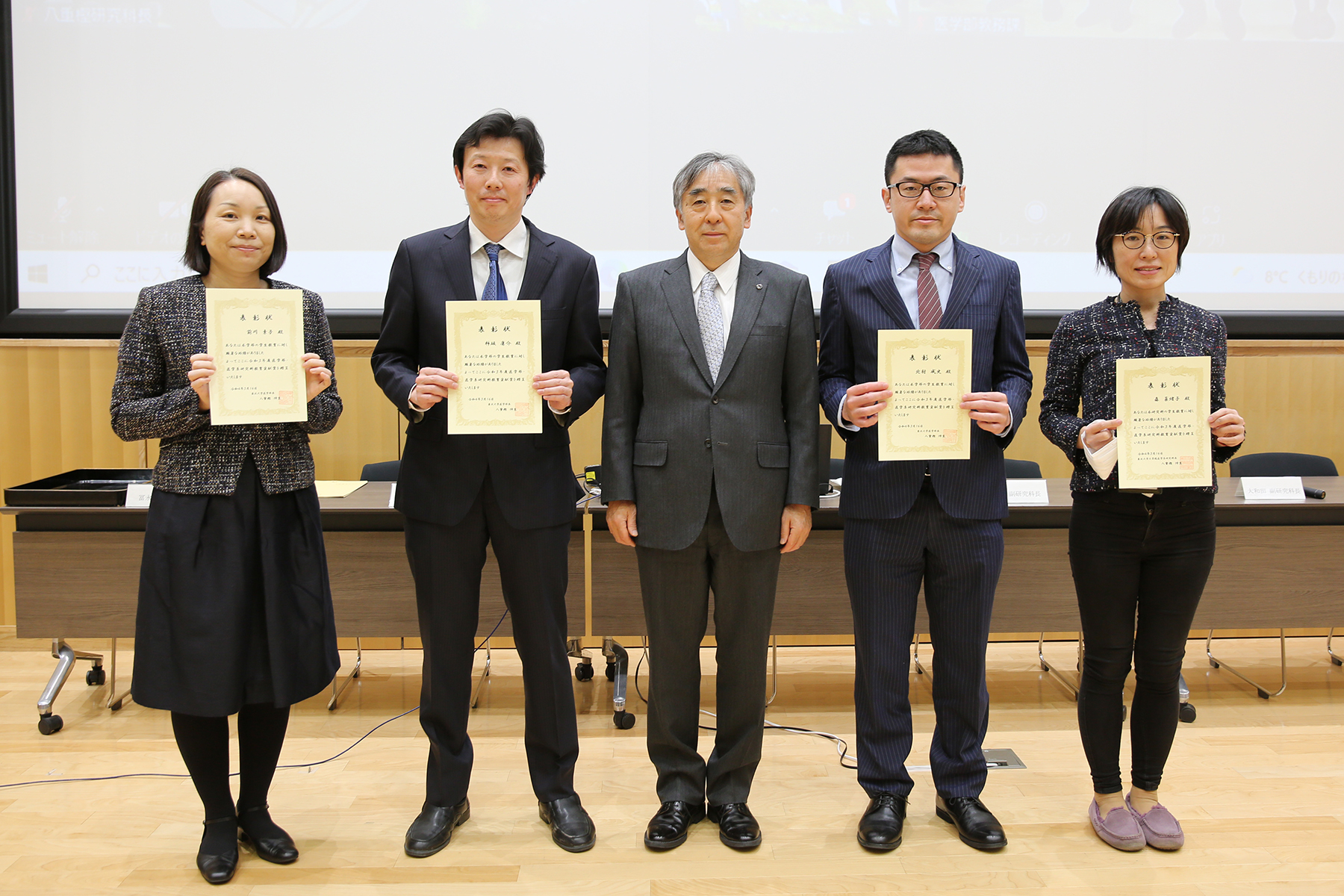 令和3年度医学部・医学系研究科教育貢献賞授与式が行われました