医科学専攻 保健学専攻
- Master's Courses
修士課程 - Doctoral Courses
博士課程
Diagnostic Imaging画像診断学
STAFF
Professor
-
Okamoto, YoshikazuProfessor. 岡本 嘉一 教授
Other Faculty / Staff
-
Kobayashi, Tomoya
Assistant Prof. 小林 智哉 助教
CONTACT
TEL:+81-22-717-7481
E-MAIL:yoshikazu.okamoto.b3*tohoku.ac.jp
(「*」を「@」に変換してください)
OUTLINE
In our laboratory,
“Diagnostic Imaging x AI x Contribution to Society = Well-being”
is the philosophy of our laboratory.
We are conducting research to help people from all walks of life to live actively in their 100-year lives through the power of diagnostic imaging. Specifically, we are conducting research on sports injury prevention for boys and girls, sports injury prevention for seniors, and upper limb checkups for wheelchair users. Some examples of our research are as follows
1) Skeletal muscle MRI
MRI has recently become able to provide not only tomographic images but also various other biological information. We are applying this information to skeletal muscle and studying its application to lipid metabolism in the human body, muscle conditioning in athletes, and indicators of rehabilitation effectiveness (Figure 1). 2.
2) Sports Examination by In-vehicle MRI
In collaboration with the University of Tsukuba, we have developed a vehicle-mounted ultra-compact MRI system and have conducted many on-site MRI sports examinations. In addition to sports examinations for boys and girls and “upper limb examinations” for wheelchair users, we plan to offer “tennis examinations” and “golf examinations” for seniors in the future.
3) Prediction of Disability Prevention by AI
Our department also conducts research on postmortem imaging. As Japan transitions from an aging society to one with a high mortality rate, accurate determination of the cause of death becomes increasingly important to maintain social order. Postmortem imaging has become a crucial tool for determining the cause of death in Japan, where the autopsy rate is low, and it is also being utilized for identifying unidentified bodies.
Our research aims to enhance the technology for determining the cause of death and identifying individuals using postmortem imaging.
4) Postmortem Imaging (Autopsy imaging)
Our department has traditionally focused on research using AI. Key to the use of medical imaging AI is the quality and quantity of images, known as “teacher data”. In addition, data on areas and attributes that are not usually captured are particularly important because of their scarcity.
We can collect a lot of data with special attributes in the above “on-site sports medical checkups” (Figs. 2 and 3). The “skeletal muscle MRI” tractography image (Figure 1) is also difficult to obtain because it requires know-how to generate. We are applying these data to AI to assist in diagnostic imaging and to predict injury prevention.
We will continue to disseminate our research results for the prevention of disabilities in youth sports, prevention of disabilities in senior sports, conditioning, and rehabilitation, so that people can “live better” using diagnostic imaging technology in the age of 100 years of life.
我々の教室では、
「画像診断×AI×社会貢献=Well-being 」
を研究室の理念として日々活動しています。
我々は画像診断の力で、様々な立場の方が人生100年時代をactive に過ごしてもらうための一助となるような研究を行っています。具体的には少年少女スポーツ障害予防、シニアスポーツ障害予防、車椅子ユーザーの上肢検診などです。その研究例を幾つか下記に示します。
1. 骨格筋MRI
近年、MRIは断層画像だけでなく、様々な生体情報を得ることができるようになっています。我々はこれらを骨格筋に応用し、人体の脂質代謝やアスリートの筋コンディショニング、リハビリテーション効果の指標(図1)等に応用する研究を行っています。
2. 車載型MRI によるスポーツ検診
筑波大学と共同で車載型超小型MRI 装置を開発し、多くの出張MRIスポーツ検診を行ってきました。少年少女スポーツ選手検診、車椅子ユーザーの「上肢検診」に加え(図2,3)、今後はシニア世代の「テニス検診」「ゴルフ検診」、を行う予定です。
3. オートプシーイメージング(死亡時画像診断)
当教室では、1~3の研究に加えて、死亡時画像診断についての研究も行っています。
日本は、高齢化社会から多死社会に向かっており、社会秩序を保つために正確な死因究明が重要になってきています。死亡時画像診断は、解剖率が低い日本における死因究明の重要なツールとして普及しており、身元不明遺体の個人特定にも活用されようとしています。
我々の研究では、死亡時画像診断を用いた死因究明および個人識別の技術の向上を目指しています。
4. AI による障害予防予測
当教室は伝統的にAIを使った研究に力を入れております。医療用画像診断AI の活用において鍵となるのは、「教師データ」と言われる画像の質と量です。また、通常は撮影されない部位や属性のデータは希少性が高いため、特に重要です。
我々は上記「出張スポーツ検診」で特殊な属性を持つデータを多く収集する事が可能です(図2,3)。「骨格筋MRI」のトラクトグラフィーという画像(図1)にも生成にノウハウが必要で入手困難です。我々はこれらのデータをAIに応用し、画像診断補助や、障害予防予測の研究に取り組んでいます。
我々は、少年少女スポーツの障害予防、シニアスポーツの障害予防、コンディショニング、リハビリテーションなど、人生100年時代に画像診断技術を用いて人々が「よりよく生きる」ための研究成果を発信していきます。
Figure 1.Diffusion tensor of MRI can be applied to depict skeletal muscle fibers. We are considering its application as a new index for rehabilitation.
図1.MRI拡散テンソルという技術を応用し、骨格筋の筋線維を描出している。 リハビリテーションの新指標としての応用を考えている。
Figure 2.Vehicle-mounted MRI is used to check-up for prevention of sports injury for kids and seniors, and upper limb of wheelchair users.
図2.超小型MRI を搭載した検診車にて、子供やシニアのスポーツ検診、車椅子ユーザーの上肢検診等を行っている。
Figure 3.An image of very early stage baseball elbow captured by a vehicle-mounted MRI. AI is applied to diagnose and predict the prognosis of these screening images.
図3.車載型MRIで超早期の野球肘を捉えた画像。これら検診画像をAIで診断し、予後を予測する研究を行っている。
Scenery of Laboratory
研究室風景
ARTICLE
Sugawara K, et al. Breast cancer classification based on breast tissue structures using the Jigsaw puzzle task in self-supervised learning. Radiological Physics and Technology. 18(1):209-218, 2025.
URL:https://doi.org/10.1007/s12194-024-00874-y
Kohyama S, et al. Traction MRI of the Elbow: Age-Based Effects and Implications. Diagnostics. 14(19):2165, 2024.
URL:https://doi.org/10.3390/diagnostics14192165
Kohyama S, et al. Optimization of Traction Magnetic Resonance Imaging to Improve Visibility of the Elbow Cartilage. Diagnostics. 14(6):630, 2024
URL:https://doi.org/10.3390/diagnostics14060630
Sado N, et al. Hip and lumbosacral joint centre locations in asian population: Biases produced by existing regression equations and development of new equations. Journal of Biomechanics. 162:111866, 2024.
URL:https://doi.org/10.1016/j.jbiomech.2023.111866
Kobayashi T, et al. Usefulness of fat attenuation index in postmortem CT for identifying responsible vessels in acute coronary syndromes: A case report. Radiology Case Reports. 19(11), 5384-5388, 2024.
URL:https://doi.org/10.1016/j.radcr.2024.08.030
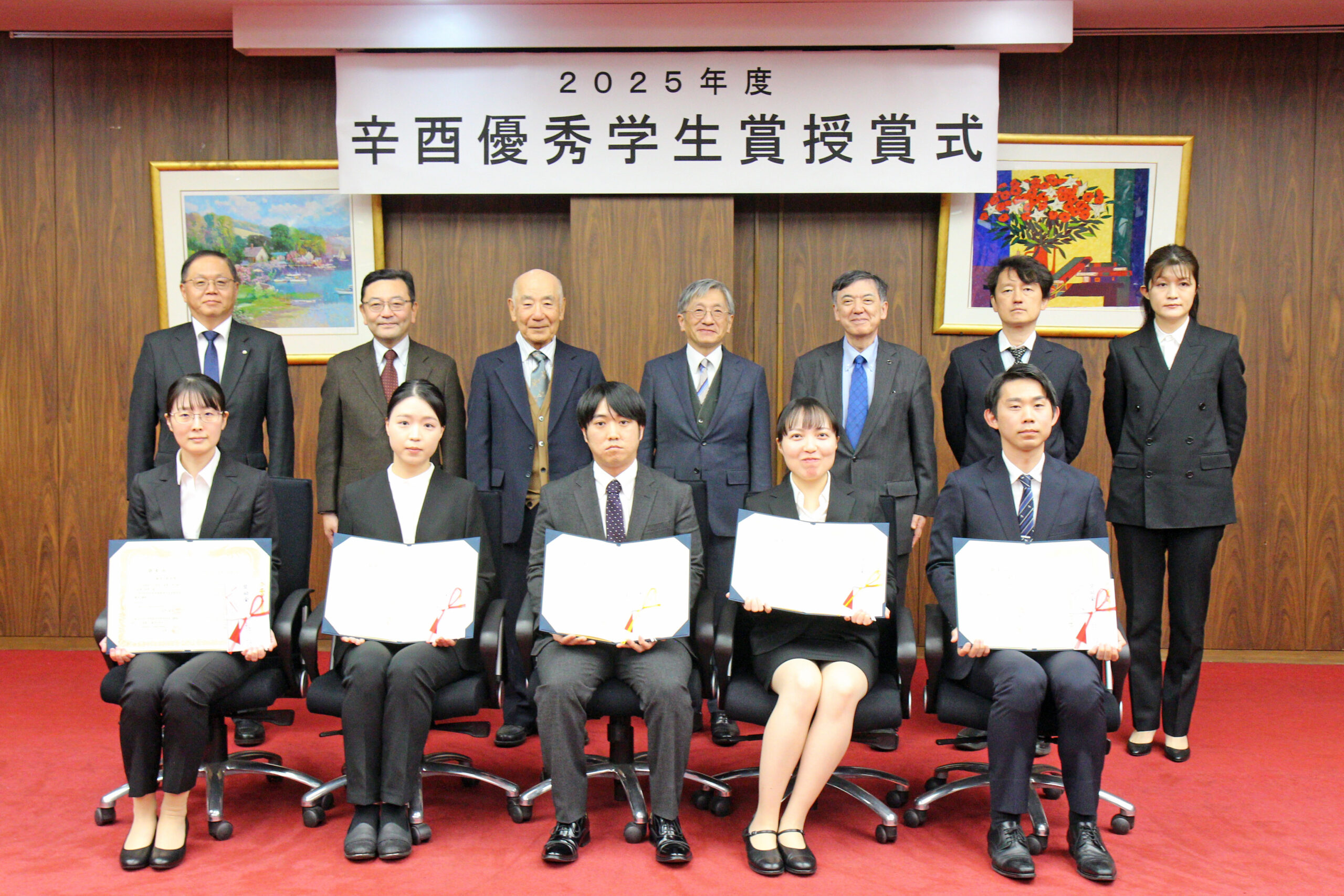 2025年度 辛酉優秀学生賞授与式を行いました
 画像診断学分野 岡本 嘉一教授の記事が東京新聞Tokyo webに掲載されました