保健学専攻
- Master's Courses
修士課程 - Doctoral Courses
博士課程
Diagnostic Image Analysis画像解析学
STAFF
Professor
-
Kaneta, TomohiroProfessor. 金田 朋洋 教授
Other Faculty / Staff
-
Usui, Akihito
Lect. 臼井 章仁 講師 -
Odagiri, Hayato
Lect. 小田桐 逸人 講師
CONTACT
TEL:+81-22-717-7872
E-MAIL:kaiseki*med.tohoku.ac.jp
(「*」を「@」に変換してください)
OUTLINE
In diagnostic imaging, we are extensively studying image analysis techniques using nuclear medicine and PET, and their application to treatment.
Imaging and therapeutic applications using nuclear medicine and PET: We are studying the standardization and optimization of imaging conditions and timing in nuclear medicine and PET. At Tohoku University, many new PET agents have been developed at the Research Center for Accelerator and Radioisotope Science (RARiS). Recently, we have been competing with the world in the development of PET agents for amyloid and tau, which are thought to be the cause of Alzheimer's disease. In December 2023, Recanemab, a drug that removes amyloid from the brain, was launched in Japan. In order to use this drug, it is essential to confirm the accumulation of amyloid in the brain by amyloid PET. We are also working on the clinical study of a new tau PET drug. In addition, we are working on “theranostics” research combining PET drugs with nuclear medicine therapy using alpha-ray emitting radionuclides, which can be provided at RARiS.
Postmortem image analysis: In cooperation with the Miyagi Prefectural Police, the Department of Forensic Medicine, and the Department of Diagnostic Radiology, we are conducting research on postmortem image diagnosis methods, focusing on CT before forensic autopsy. We are creating 3D images of bones from postmortem CT images, investigating the frequency of normal variations, and studying methods for determining gender and estimating age using various measurements of bones. We are researching methods for diagnosing lesions in postmortem images based on the weight and CT values of various organs.
画像診断の中でも核医学、PETを用いた画像解析技術、治療への応用を幅広く研究しています。
1) 核医学、PETを用いた画像診断、治療への応用:核医学、PETにおける撮影条件や画像の標準化・最適化を研究しています。また東北大学では先端量子ビーム科学研究センター(旧:サイクロトロン・RIセンター)にて多くの新規PET薬剤を開発しています。最近ではアルツハイマー病の原因と考えられているアミロイドやタウのPET薬剤開発で、世界と競合してきました。2023年12月にアルツハイマー病の疾患修飾薬レカネマブが我が国で発売となりましたが、この薬剤を使用するためにはアミロイドPETで脳内のアミロイド蓄積を確認することが必須です。また新たな第二世代タウPET薬剤の臨床研究実施に向けて、現在取り組んでいます。さらに先端量子ビーム科学研究センターで供給可能なα線放出核種を用いた核医学治療とPET薬剤を組み合わせたセラノスティクス(Theranostics)研究も進めていきます。
2)死後画像解析:宮城県警、法医学分野、画像診断学分野と協力して法医解剖前CTを中心とした死後画像診断法の研究を行っています。死後CT画像から骨の3次元画像を作成し、破格の頻度の調査や、骨の各種計測による性別判定や年齢推定法を研究しています。各種臓器の重量やCT値などから死後画像における病変の診断法を研究しています。
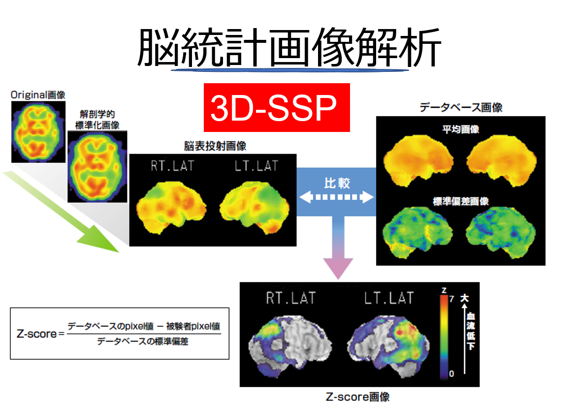
脳血流SPECTの統計画像解析(3D-SSP)のスキーム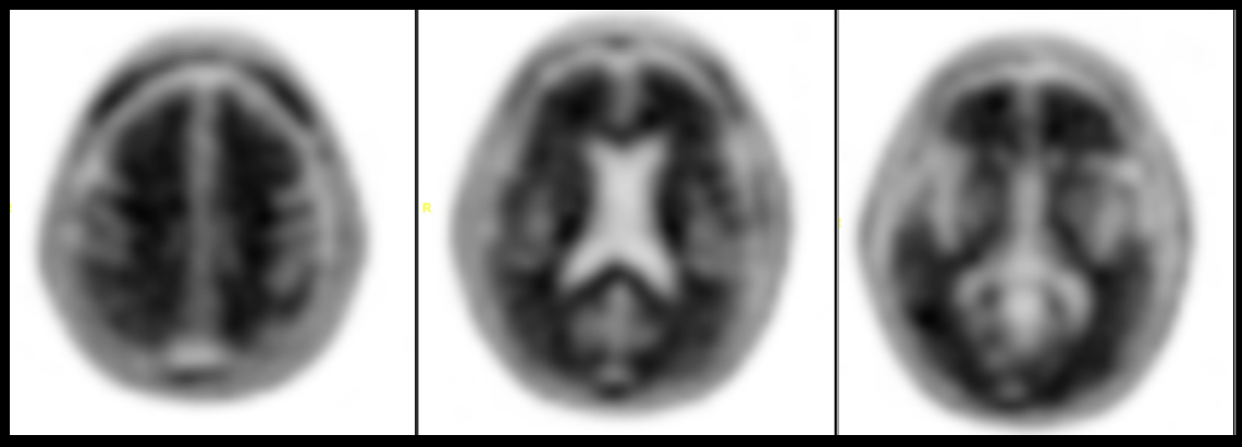
アミロイドPET陽性像
ARTICLE
Odagiri H, et al. Verification of the effect of data-driven brain motion correction on PET imaging. PLoS One. 2024 Jul 5;19(7):e0301919. doi: 10.1371/journal.pone.0301919. eCollection 2024.
Sasaki Y, et al. A case report of forensic personal identification by transposed teeth and dental treatment marks using multidetector row computed tomography. Radiol Case Rep. 2023 Nov 23;19(2):581-585.
Odagiri H, et al. Improvement of Imaging Conditions to Improve the Detection Rate of Head and Neck Cancer by Positron Emission Tomography/Computed Tomography Examination. Tohoku J Exp Med. 2023 May 30;260(2):141-147.
Okumura S, et al. Diagnostic Accuracy of Liver Damage Based on Postmortem Computed Tomography Findings in High-Energy Trauma. Tohoku J Exp Med. 2022 Jul 22;257(4):327-332.
Sato K, et al. Accuracy of virtual monochromatic images generated by the decomposition of photoelectric absorption and Compton scatter in dual-energy computed tomography. Phys Eng Sci Med. 2022 Mar;45(1):239-249.
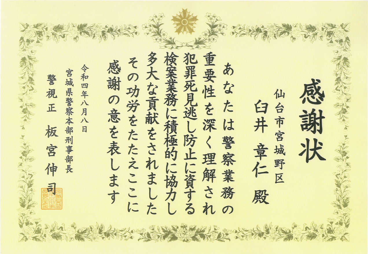 画像解析学分野/Aiセンターの臼井章仁先生が、宮城県警察本部より感謝状を授与されました。