その他
Ophthalmic Imaging and Information Analytics眼科画像情報解析学
STAFF
Professor
-
Nakazawa, ToruProfessor. 中澤 徹 教授 (兼任)

Other Faculty / Staff
-
Ishikawa, Makoto
Assoc. Prof. 石川 誠 准教授
CONTACT
TEL:+81-22-717-7294
E-MAIL:eye*oph.med.tohoku.ac.jp
(「*」を「@」に変換してください)
OUTLINE
Glaucoma is a disease in which the optic nerve is damaged and the visual field is lost. Due to the irreversible nature of the change, the lost field of vision is irreversible. It is important to diagnose glaucoma at an early stage, determine its progression, and provide treatment that is appropriate for each individual case.
We have clarified the relationship between retinal thickness and visual acuity using optical coherence tomography (OCT). This achievement has enabled us to recognize cases of vision loss at an early stage in daily practice (Ref. 1).
Glaucoma is a multifactorial disease, and there are cases in which visual field damage progresses even after treatment has sufficiently lowered intraocular pressure. Glaucoma is a multifactorial disease, and the quality of medical care can be improved by segmentation. One of the risk factors is decreased ocular blood flow. We used OCT to identify the characteristics of patients with reduced ocular blood flow (Ref.2,3). This is an important topic that will lead to individualization of treatment.
We are conducting advanced research to utilize accumulated medical knowledge for diagnosis and segmentation using imaging technology and artificial intelligence (Ref. 4, 5). From big data, we are preparing to predict prognosis and provide preemptive medicine by making full use of biomarkers such as imaging data, genes, clinical background, and oxidative stress. Let's work together to advance research toward the goal of zero blindness.
緑内障は視神経が障害を受け、視野が欠けていく疾患です。不可逆性の変化のため、失われた視野は元に戻すことが出来ません。早期の診断・進行判定、個々の症例に適した治療が重要です。
QOLには良好な視力が大事ですが、我々は光干渉断層計(Optical coherence tomography; OCT)を用いて網膜厚と視力の関係を明らかにしました。この成果により、日常診療でも視力低下を来す症例に、早期に気づけるようになりました(文献1)。
緑内障は治療によって眼圧下降が十分に得られても、視野障害が進行する症例が存在し、現状では治療が十分とは言えません。緑内障は多因子疾患であり、細分化により診療の質が上がります。危険因子の一つに眼血流低下があります。我々はOCTを用いて、眼血流低下する症例の特徴を明らかにしました(文献2,3)。治療の個別化につながる重要なテーマです。
積み重ねられた医療の知見を、画像技術と人工知能を駆使し、診断や細分化に役立てる先進的な研究を行っています(文献4,5)。ビッグデータから、画像データ・遺伝子・臨床背景・酸化ストレスなど、最先端のバイオマーカーを駆使して、予後を予測し先制医療を行う準備を進めています。失明ゼロを目指し、ともに研究を進めましょう。
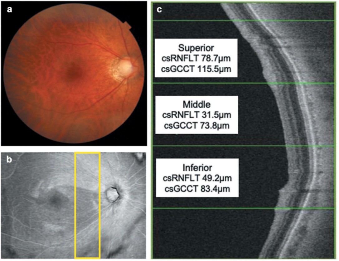
Relationship between papillomacular nerve fiber bundle and visual acuity by OCT (Ref.1)
OCT画像による乳頭黄斑線維と視力の関連 (文献1)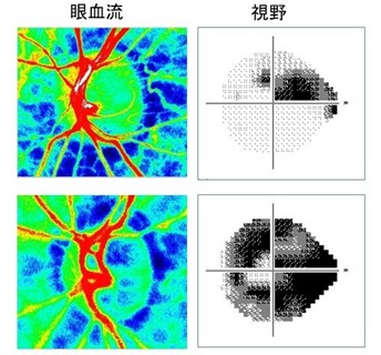
Examples of ocular blood flow and visual field defect and correction of ocular blood flow (Ref. 2)
眼血流と視野障害の例と眼血流の補正 (文献2)
ARTICLE
Takahashi N, et al. Evaluation of Papillomacular Nerve Fiber Bundle Thickness in Glaucoma Patients with Visual Acuity Disturbance. Curr Eye Res. 45(7): 847-853, 2020.
URL:https://pubmed.ncbi.nlm.nih.gov/31880172/
Omodaka K, et al. Structural Characterization of Glaucoma Patients with Low Ocular Blood Flow. Curr Eye Res. 45(10): 1302-1308, 2020
URL:https://pubmed.ncbi.nlm.nih.gov/32134693/
Kiyota N, et al. Time-course Changes in Optic Nerve Head Blood Flow and Retinal Nerve Fiber Layer Thickness in Eyes with Open-angle Glaucoma. Ophthalmology. 128(5): 663-671, 2021.
URL:https://pubmed.ncbi.nlm.nih.gov/33065167/
An G, et al. Glaucoma Diagnosis with Machine Learning Based on Optical Coherence Tomography and Color Fundus Images. J Healthc Eng. Feb 18: 4061313, 2019.
URL:https://pubmed.ncbi.nlm.nih.gov/30911364/
An G, et al. Hierarchical deep learning models using transfer learning for disease detection and classification based on small number of medical images. Sci Rep. 11(1):4250, 2021.
URL:https://pubmed.ncbi.nlm.nih.gov/33649375/