医科学専攻
- Master's Courses
修士課程 - Doctoral Courses
博士課程
Advanced Neuological Surgery先進脳神経外科治療学講座(広南会広南病院)
STAFF
Professor
-
Omodaka, ShunsukeProfessor. 面高 俊介 教授
CONTACT
TEL:+81-22-248-2131
E-MAIL:shunsuke.omodaka.e1*tohoku.ac.jp
(「*」を「@」に変換してください)
OUTLINE
It is our distinct pleasure to welcome all the young investigators, being interested in the cerebrovascular surgeries for intracranial aneurysms, Moyamoya disease, and brain vascular malformations. We are particularly focusing our interests on the basic pathology and perioperative cerebral hemodynamics in patients with Moyamoya disease. Cerebral blood flow measurement after revascularization surgery for Moyamoya disease significantly contributed to the understanding of the intrinsic and complex perioperative pathology of Moyamoya disease, and has markedly improved the outcome of surgery during the past 10 years. We are also focusing on the basic pathology of intracranial aneurysms and their growth/rupture using high-resolution MRI vessel wall imaging technique, which is now clinically applied to evaluate the risk of future aneurysm growth and/or rupture in each patient. We are also conducting clinical/basic research of dissecting aneurysms and brain vascular malformations.
当科では、近年発展が目覚しい脳血管障害に対する外科治療を入口として、脳動脈瘤、もやもや病、脳血管奇形などの脳血管疾患における新規治療開発・病態研究・脳循環代謝研究を行っています。特に、若年者脳卒中の原因として近年増加傾向にあるもやもや病に関しては半世紀以上にわたる当科での診療経験を踏まえて、遺伝子研究やバイパス手術に際しての周術期病態の解明を中心に先進的研究を推進しています。また、くも膜下出血の原因として重要な脳動脈瘤に関しては高感度MRIを用いた動脈瘤壁イメージング(vessel wall imaging)やコンピューター流体解析(computational fluid dynamics)を駆使した動脈瘤増大・破裂に関する病態研究を進めています。
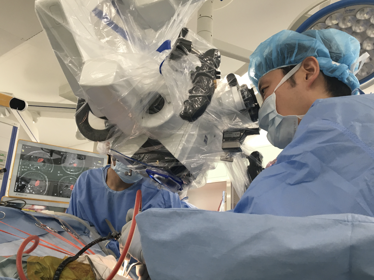
Intraoperative photo of bypass surgery for Moyamoya disease
もやもや病に対するバイパス術中写真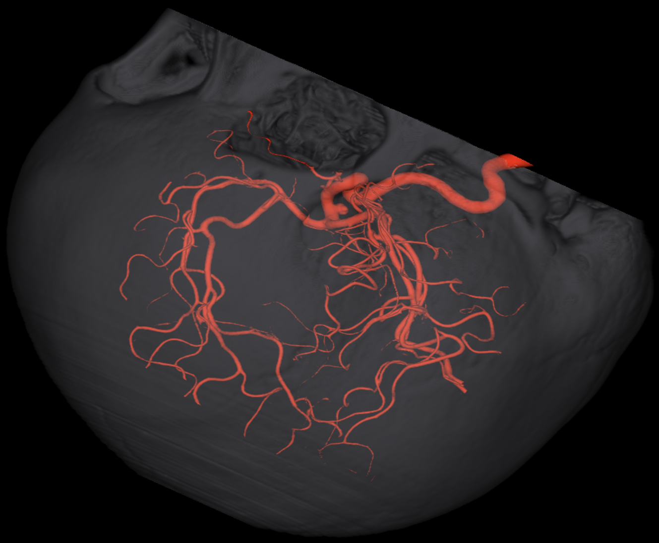
Intraoperative photo of clipping surgery for intracranial aneurysm
脳動脈瘤に対するクリッピング術の術中写真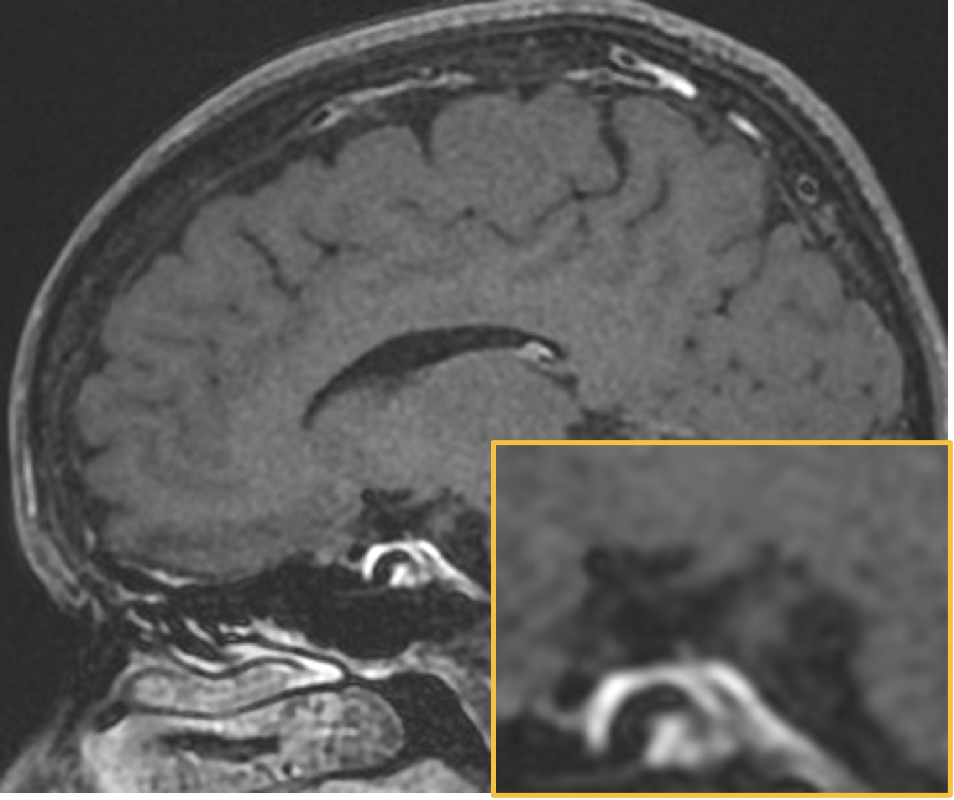
Intraoperative photo of clipping surgery for intracranial aneurysm
脳動脈瘤に対するクリッピング術の術中写真
ARTICLE
Omodaka S, Endo H , et al. Aneurysm Wall Enhancement Can Predict Rupture Point in Intracranial Aneurysms With Multiple Blebs. Neurosurgery. 96(3) 593-599 2025
Omodaka S, Endo H , et al. Six-month Outcomes after PulseRider- and Conventional Single Stent-assisted Embolization for Bifurcation Aneurysms: A Propensity-adjusted Comparison. Neurol Med Chir (Tokyo). 63(11) 512-518 2023
Omodaka S, Endo H , et al. Wall enhancement in unruptured posterior communicating aneurysms with oculomotor nerve palsy on magnetic resonance vessel wall imaging. J Neurosurg. 137(3) 668-674 2022
Omodaka S, Endo H , et al. Circumferential wall enhancement in evolving intracranial aneurysms on magnetic resonance vessel wall imaging. J Neurosurg. 131(4) 1262-1268 2019
Omodaka S, Endo H , et al. Circumferential Wall Enhancement on Magnetic Resonance Imaging is Useful to Identify Rupture Site in Patients with Multiple Cerebral Aneurysms. Neurosurgery. 82(5) 638-644 2018
Omodaka S, Endo H , et al. Quantitative Assessment of Circumferential Enhancement along the Wall of Cerebral Aneurysms Using MR Imaging. AJNR Am J Neuroradiol. 37(7) 1262-6 2016
Omodaka S, Endo H , et al. High-grade Cerebral Arteriovenous Malformation Treated with Targeted Embolization of a Ruptured Site: Wall Enhancement of an Intranidal Aneurysm as a Sign of Ruptured Site. Neurol Med Chir (Tokyo). 55(10) 813-7 2015
Omodaka S, et al. Local hemodynamics at the rupture point of cerebral aneurysms determined by computational fluid dynamics analysis. Cerebrovasc Dis. 34(2) 121-9 2012