医科学専攻 障害科学専攻
- Master's Courses
修士課程 - Doctoral Courses
博士課程
Ophthalmology眼科学
STAFF
Professor
-
Nakazawa, ToruProfessor. 中澤 徹 教授
Other Faculty / Staff
-
Kunikata, Hiroshi
Assoc. Prof. 國方 彦志 准教授
CONTACT
TEL:+81-22-717-7294
E-MAIL:eye-oph*grp.tohoku.ac.jp
(「*」を「@」に変換してください)
OUTLINE
Glaucoma is one of the most important causes of blindness in Japan. Prevention of blindness due to glaucoma is an important theme aiming for a vibrant aging society.
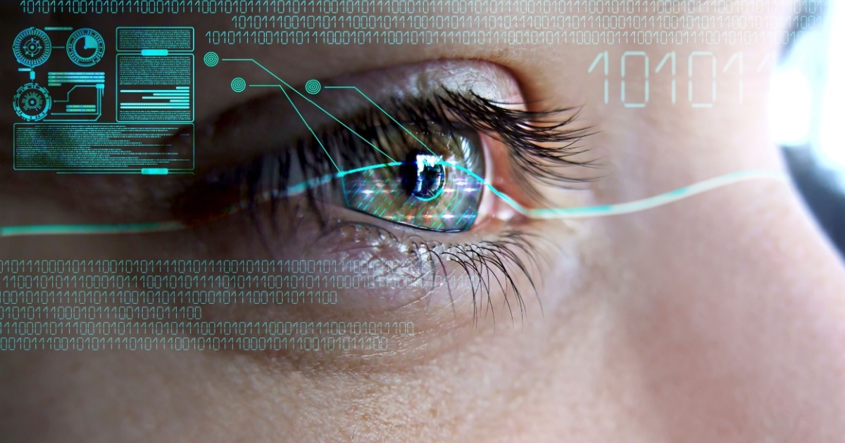
Therefore, we are working to elucidate the pathophysiology of glaucoma and develop neuroprotective treatments that suppress the progression of glaucoma. In addition, we are investigating and developing diagnostic tools that make full use of imaging technology and artificial intelligence, and conducting research to predict prognosis by using cutting-edge biomarkers such as image data, genes, clinical background, and oxidative stress from big data.
The ophthalmology department has endowed chairs called Retinal Disease Control course, Opthalmic Imaging and Information Analytics course and Advanced Ophthalmic Medicine course. Collaborating with these donated courses, we conduct research on the promotion of applications, the subdivision of glaucoma by image analysis, and the elucidation of the pathophysiology and make efforts to develop advanced medical care that integrates basic research and clinical research. For this reason, our ophthalmology department has a wide variety of work contents, so we are looking for human resources who are familiar with a work such as data analysis, laboratory assistants, patient nursing, and assistance, not limited to doctors.
緑内障は我が国の失明原因第1位となっています。緑内障による失明の予防は、活気ある高齢社会を目指すために重要なテーマです。
そのため我々は、緑内障の病態を解明し、緑内障の進行を抑制する神経保護治療の開発に取り組んでいます。また、その成果を基盤としてドラッグスクリーニングシステムによる臨床応用可能な神経保護薬の探索・開発を行っています。加えて、画像技術と人工知能を駆使した診断ツールの開発や、ビッグデータから、画像データ・遺伝子・臨床背景・酸化ストレスなど、最先端のバイオマーカーを駆使して、予後を予測する研究を行っています。
これらの研究成果を臨床で実践するトランスレショナルリサーチを目標としています。
また、当眼科学教室には網膜疾患制御学寄附講座、視覚先端医療学寄附講座および眼科画像情報解析学寄付講座があり、それぞれ網膜疾患に対する先進医療の開発、網膜変性対する治療法の開発と臨床応用の推進、画像解析による緑内障の細分化と病態解明の研究を行なっております。これらの寄付講座とも連携することで基礎研究と臨床研究を融合させた先進的医療の発展に尽力しています。
このように当教室では業務内容が広く多岐にわたるため、医師に限らず、データ解析・実験助手・患者看護・補助など多種多様な業務に精通した人材を求めています。
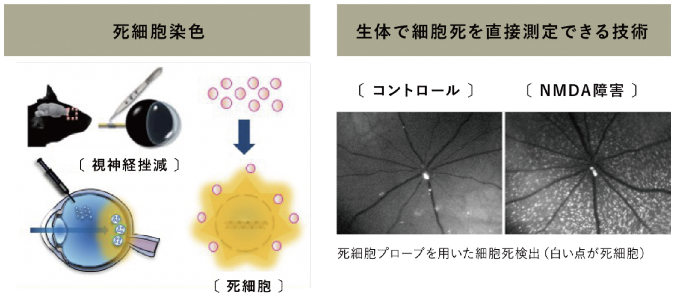
Development of in vivo imaging for retinal ganglion cell death
網膜神経節細胞死のin vivoイメージング法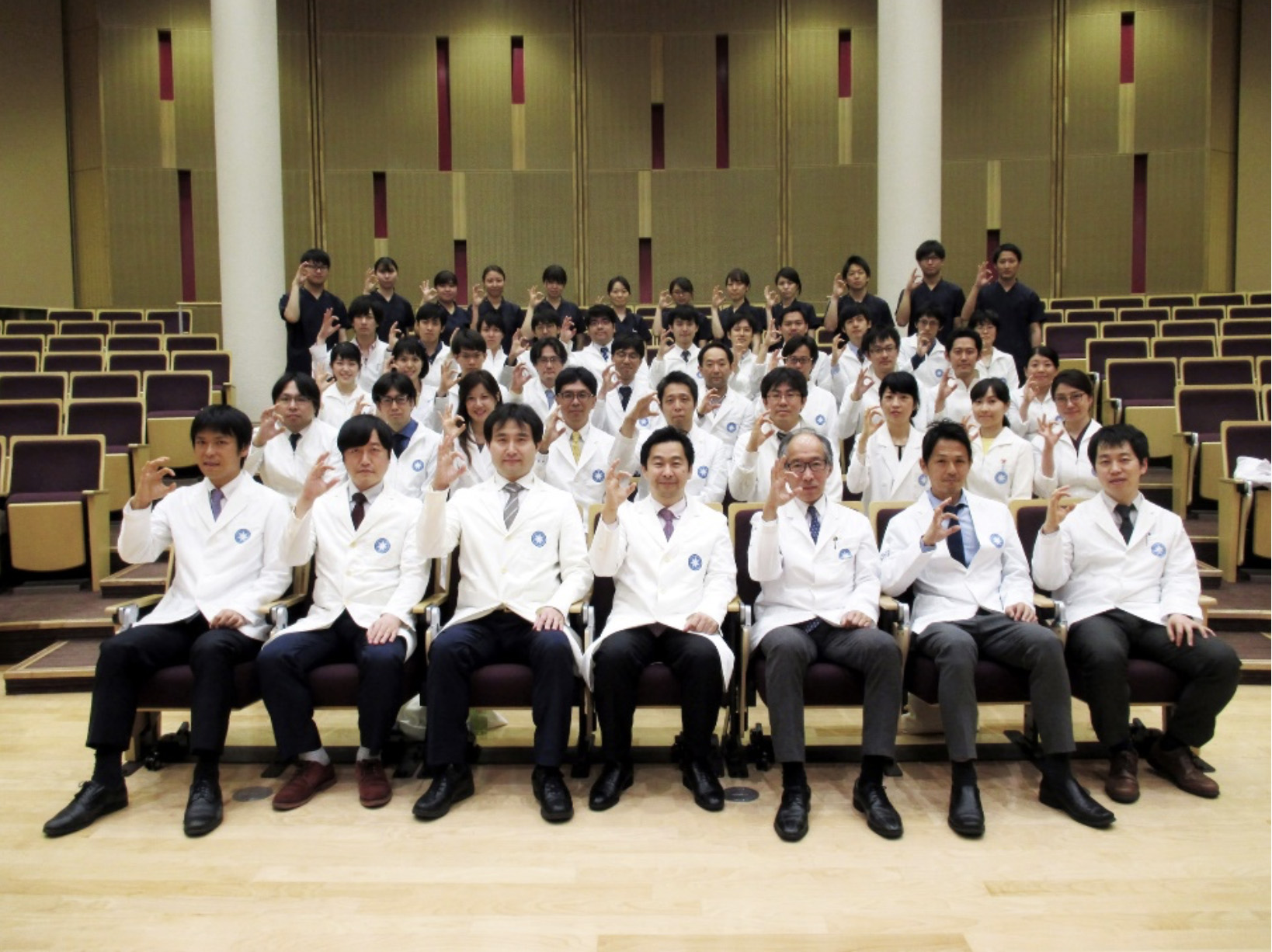
Group photo of staff members
医局員全員の集合写真
ARTICLE
Takahashi N, et al. ENO1 Dysfunction-Mediated Glycolytic Attenuation Exacerbates Oxidative Stress-Induced Retinal Ganglion Cell Death via Altered ATP Synthesis Pathway. Invest Ophthalmol Vis Sci. 66(9):63, 2025
URL:https://doi.org/10.1167/iovs.66.9.63
Sharma P, et al. A hybrid multi model artificial intelligence approach for glaucoma screening using fundus images. NPJ Digit Med. 8(1):130, 2025
URL:https://doi.org/10.1038/s41746-025-01473-w
Akiyama M, et al. Genetic Risk Stratification of Primary Open-Angle Glaucoma in Japanese Individuals. Ophthalmology. 131(11):1271-1280, 2024
URL:https://doi.org/10.1016/j.ophtha.2024.05.026
Sato K, et al. The GPR84 molecule is a mediator of a subpopulation of retinal microglia that promote TNF/IL-1α expression via the rho-ROCK pathway after optic nerve injury. Glia. 71(11):2609-2622, 2023
URL:https://doi.org/10.1002/glia.24442
Kiyota N, et al. Time-Course Changes in Optic Nerve Head Blood Flow and Retinal Nerve Fiber Layer Thickness in Eyes with Open-angle Glaucoma. Ophthalmology. 128(5):663-671, 2021
URL:https://doi.org/10.1016/j.ophtha.2020.10.010
 1枚の眼底写真から体の年齢を示す「網膜年齢」を推定するAIを開発- 糖尿病・心疾患・脳卒中との関連を確認 -
 AI搭載のポータブル眼科検査システムを開発― 場所を選ばない眼科スクリーニングにより白内障などの早期発見へ ―
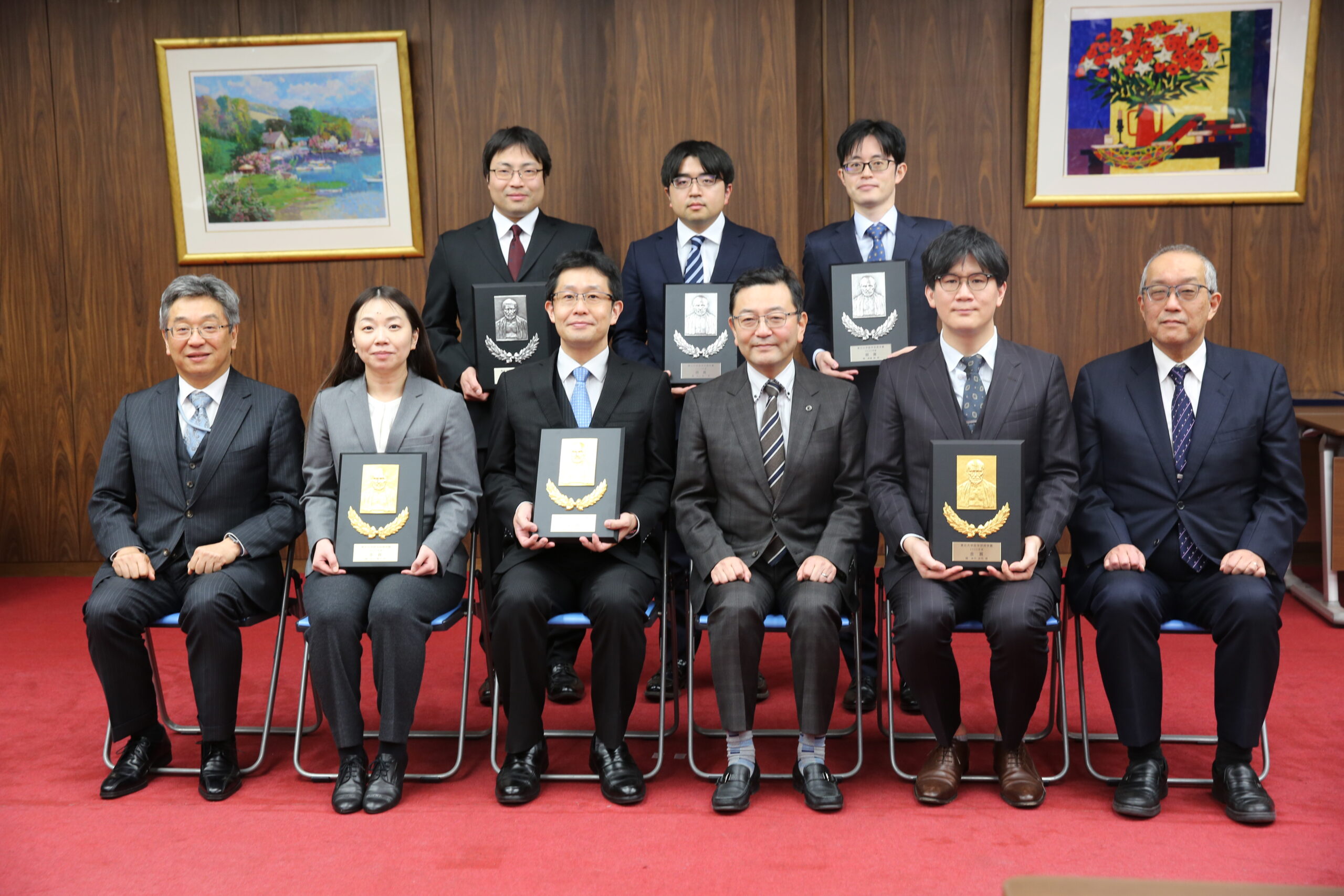 2025年度 医学部奨学賞・東北医学会奨学賞・医学部学生奨学賞授与式を行いました
 網膜中心動脈閉塞症の新たな治療薬開発へ ―医師主導治験でカルパイン阻害薬SJP-0008の安全性と有効性を確認―