医科学専攻 保健学専攻
- Master's Courses
修士課程 - Doctoral Courses
博士課程
Radiological Imaging and Informatics医用画像工学
STAFF
Professor
-
Homma, NoriyasuProfessor. 本間 経康 教授
Other Faculty / Staff
-
Ichiji, Kei
Lect. 市地 慶 講師
CONTACT
TEL:+81-22-717-8190
E-MAIL:secretary.rii.med*grp.tohoku.ac.jp
(「*」を「@」に変換してください)
OUTLINE
Radiological images acquired by using X-ray CT, fluoroscopy, and MRI can visualize what happens in the body and play important roles to achieve better diagnosis and treatment. To derive clinically useful information and findings from those radiological images, our department aims at studying and developing new methods and theories of computational intelligence including AI and machine learning technologies and implementing them as intelligent medical systems to advance the clinical diagnosis and treatment further. For example, we are developing intelligent systems that automatically detect lesions latently captured in the medical diagnosis images and classify them based on more clear explanations associated with medical evidence and practices in specialists’ diagnoses. The mathematical and computational methods to accurately track and predict the time-varying location and shape of tumors obscurely captured in X-ray images is another research topic for achieving more accurate radiation therapy. Also, we are actively advancing research collaboration with companies in the field of medical systems and making contributions to our society by releasing our developed technologies as intellectual properties.
X線CT、X線透視やMRIといった医療機器によって撮像される医用画像は、直接目にすることのできない体内の様子を可視化し、より正確な診断・的確な治療を実現する重要な情報源です。医用画像工学分野では、医用画像に含まれる有用な情報・知見を引き出すため、AI・機械学習を含む計算知能(computational intelligence)を活用した新たな理論・技術を探究し、より高度な診断・治療を支援するシステムとして実装する研究に取り組んでいます。たとえば、画像診断支援のため、医用画像に潜む病変や病因を深層学習モデルによって自動検出・識別し、くわえてその根拠を専門医のもつ高度な読影論理や画像解剖学的知見とも関連付けて提示する手法や、がん放射線治療の効果向上・副作用低減のため、X線透視像から腫瘍の位置・形状の時間変化を追跡し、予測する数理的手法などの研究・開発を行っています。また、医療用機器・システムの開発企業との共同研究も積極的に推進しており、開発した技術の知財化と移転を通して研究成果の社会還元も図っています。
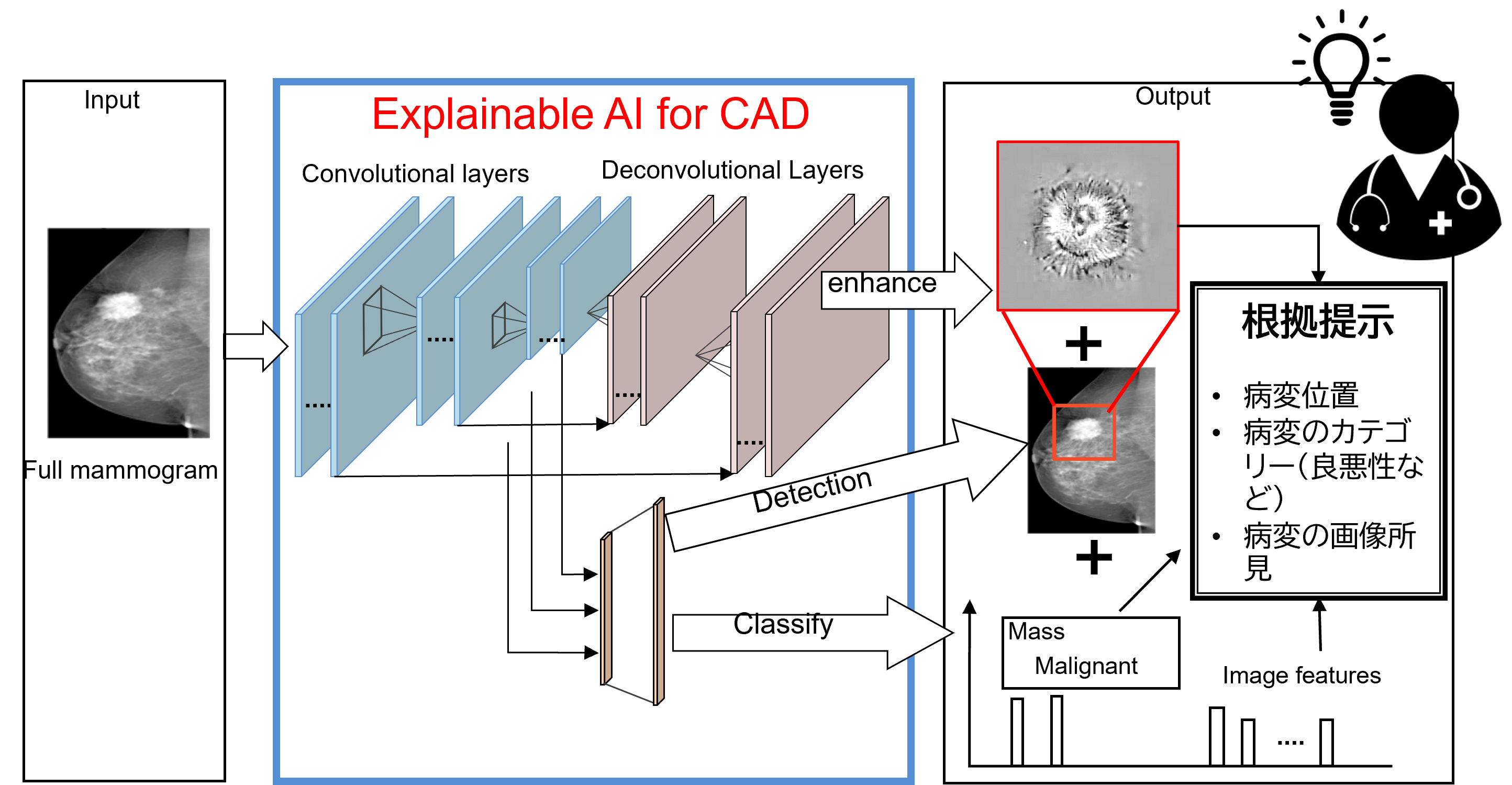
Computer aided diagnosis (CAD) system based on explainable AI
診断根拠を提示可能な画像診断支援システム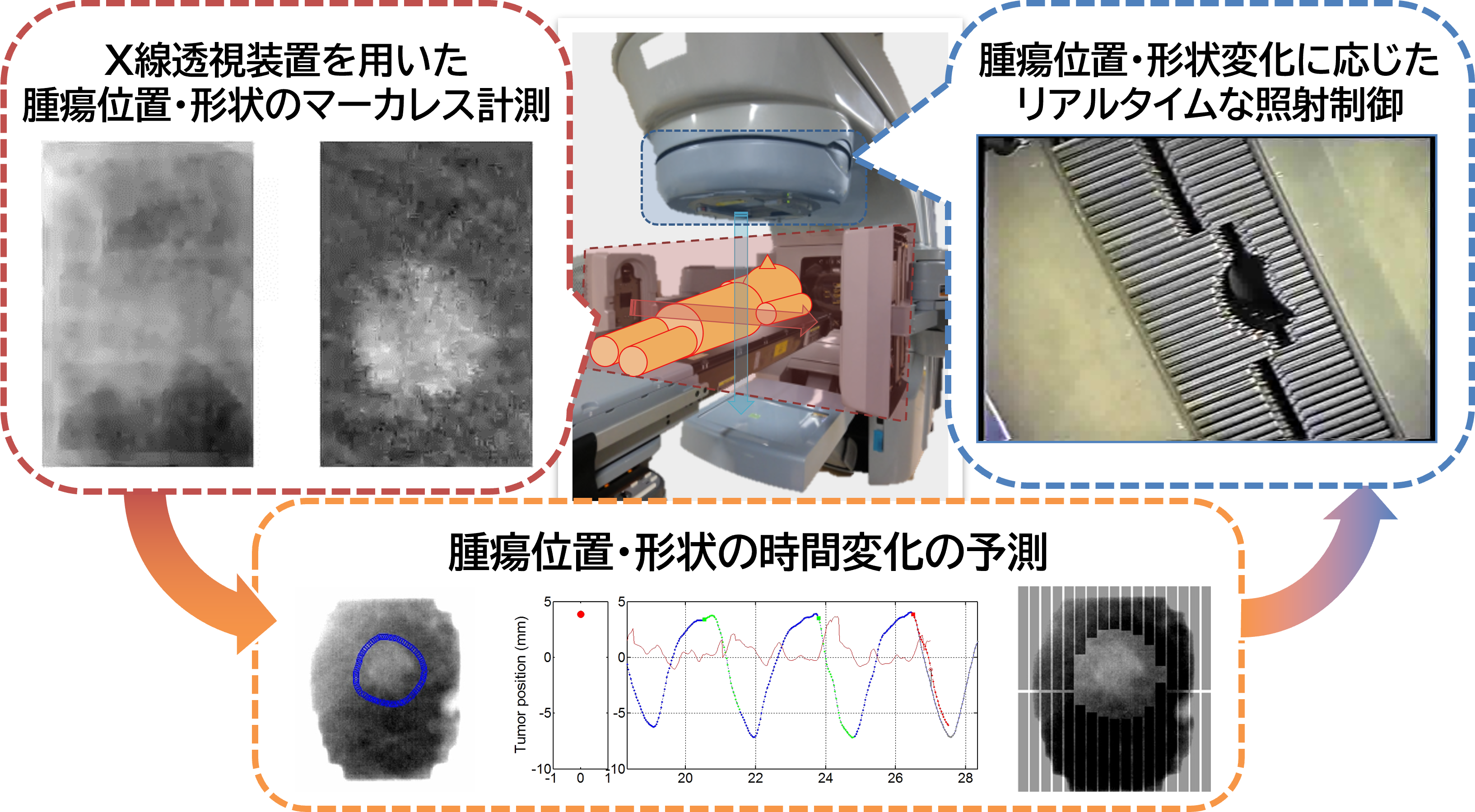
Tumor motion measurement, prediction, and control technologies for radiation therapy
放射線治療のための計測・予測・制御技術開発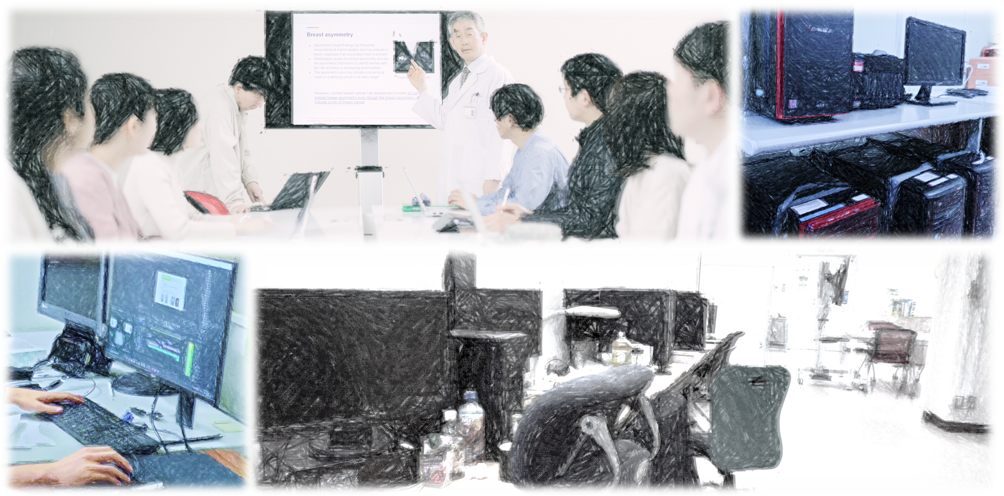
Research on medical computational intelligence by collaborating medicine and engineering
医工連携で医用システム向け計算知能を探究
ARTICLE
Bukovsky I, Dohnal G, Benes Peter M, Ichiji K, Homma N. Letter on Convergence of In-Parameter-Linear Nonlinear Neural Architectures With Gradient Learnings, IEEE Transactions on Neural Networks and Learning Systems 1-4,2021
URL:https://doi.org/10.1109/TNNLS.2021.3123533
Hoyoshi K, Ohmura T, Kayano S, Goto M, Muramatsu S, Homma N.A Review of Current Knowledge for X-ray Energy in CT: Practical Guide for CT Technologist.Nihon Hoshasen Gijutsu Gakkai zasshi,Vo.78 5 449-463,2022 [Jpn]
URL:https://doi.org/10.6009/jjrt.2022-1238
Shinohara T, Ichiji K, Wang J, Homma N, Zhang X, Sugita N, Yoshizawa M.Improved Tumor Image Estimation in X-ray Fluoroscopic Images by Augmenting 4DCT Data for Radiotherapy.Journal of Advanced Computational Intelligence and Intelligent Informatics (JACIII),Vol.26 No.4,471-482,2022
URL:https://doi.org/10.20965/jaciii.2022.p0471
Yuwen Z, Zhang X, Kawasumi Y, Usui A, Ichiji K, Funayama M, Homma N.A 2.5D Deep Learning-Based Method for Drowning Diagnosis Using Post-Mortem Computed Tomography. IEEE journal of biomedical and health informatics,Vol.27,Issue:2,1026-1035,2023
URL:https://doi.org/10.1109/JBHI.2022.3225416
Yuwen Z, Xiaoyong Z, Yoshizumi I, Zhang Z, Mizuno T, Sakamoto S, Kawasumi Y, Usui A, Ichiji K, Bukovsky I, Funayama M, Homma N. Deep Learning-Based Diagnosis of Fatal Hypothermia Using Post-Mortem Computed Tomography
The Tohoku Journal of Experimental Medicine,2023.J041,2023
URL:https://doi.org/10.1620/tjem.2023.J041
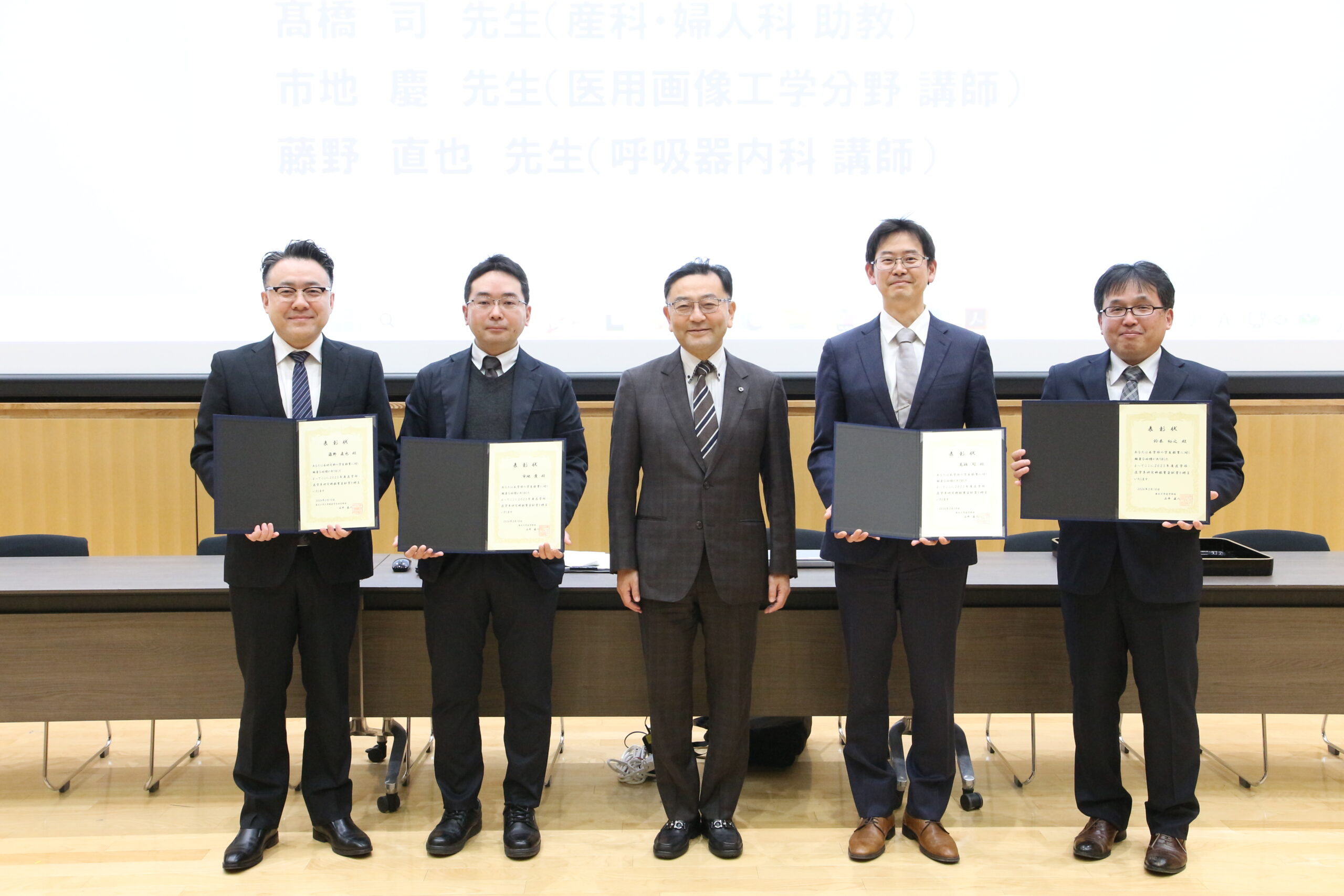 2025年度 医学部・医学系研究科教育貢献賞授与式を行いました
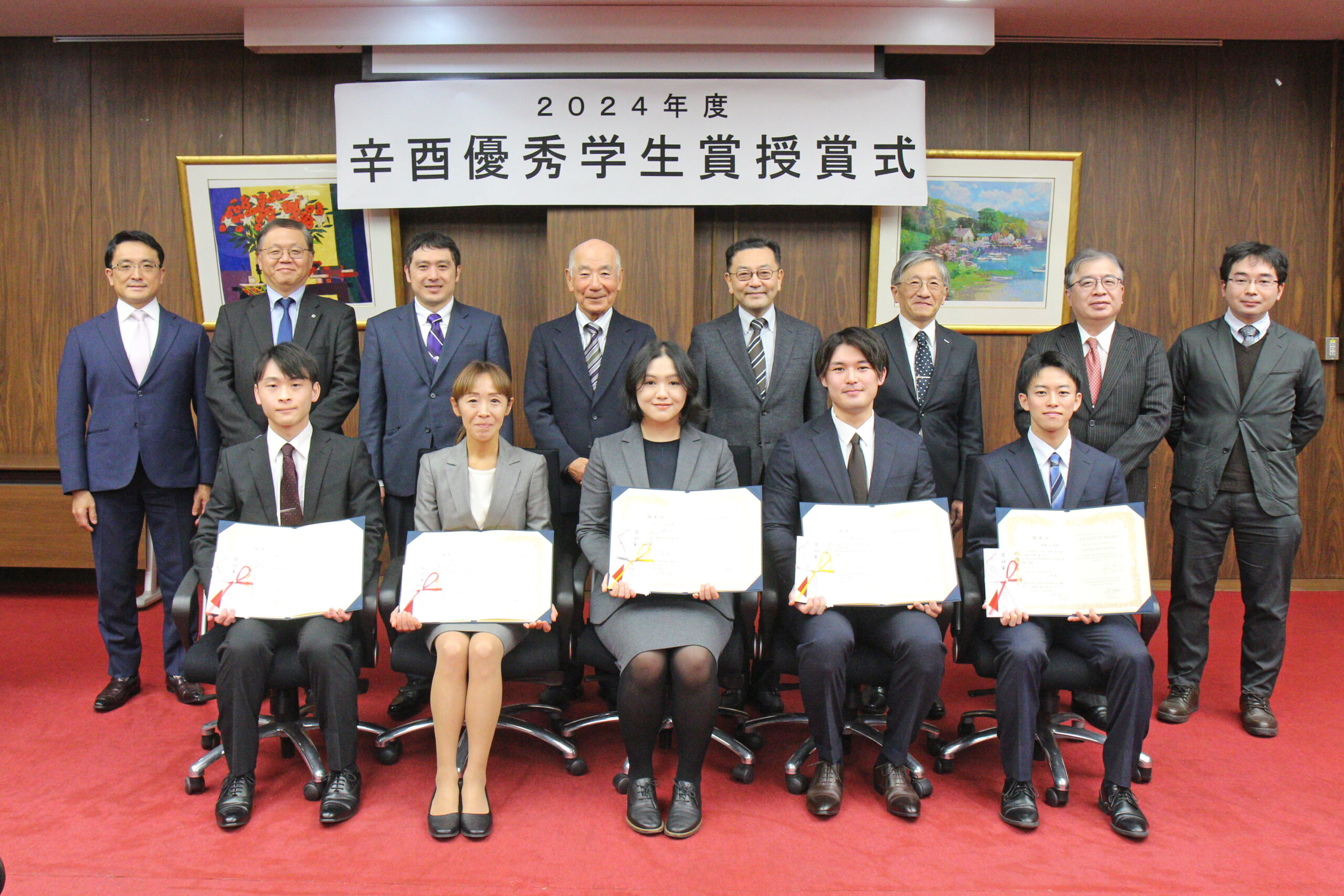 「2024年度 辛酉優秀学生賞」授賞式を行いました
 人工知能の高性能に潜む新たな危険性を解明ー医用画像診断用途の信頼性向上に期待ー